MOOC: Instrumental analysis of cultural heritage objects
2.3. Fluorescence microscopy
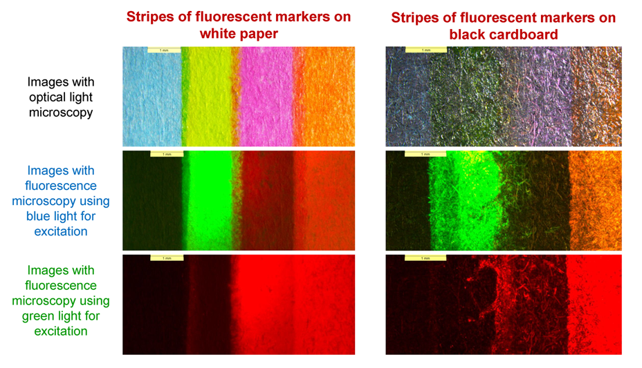
Fluorescence microscopy is another type of light microscopy that can be used to observe the samples (or particular components in the sample) that fluoresce (see Fig. 1). Fluorescence is a property of some atoms and molecules to absorb light of some specific wavelengths (used to excite the fluorescent molecules) and then promptly emit light at another, longer wavelength (this light is collected and used to form an image). Usually, fluorescence microscopy accessories are integrated with regular optical microscopes since the fluorescence instrument only needs an excitation source and additional filters. For a more detailed description of the parts of the fluorescence microscope, please see the video.
Most compounds do not fluoresce. The substance has to have a special fluorophoric structure for fluorescence emission (usually, the molecules of these compounds consisting of several aromatic rings). Many organic dyes are fluorescent (for example, hematein in logwood, carminic acid in cochineal etc.) and thus can be examined with a fluorescence microscope. In cultural heritage research, dye analysis is usually made for clothes, carpets and other textiles. Dyed textiles can be placed directly under a fluorescence microscope and observe which dyes fluoresce and at what wavelength and with what intensity. This approach can give preliminary information about the dye without sampling and extraction of the dye from the fibre (which is necessary for chromatographic analysis, see sections 5.1. and 6.1.).
Fluorescence microscopy could also be a useful tool for examining pigments in paint or detecting varnish on the paint layer. Some inorganic pigments fluorescence – zinc white has a strong yellowish fluorescence, cadmium sulphides, which fluoresce in the yellow to the red part of the spectrum, also some lake pigments and some coals fluoresce. [17]

Please see also an example of an analysis of dyed fibres with fluorescence microscopy in the video in the section of Optical microscopy.


