MOOC: Instrumental analysis of cultural heritage objects
2.2. Polarized light microscopy
Polarized light microscopy is a contrast-enhancing technique that can be used to analyse the structure of the object. Polarized light microscopes have higher sensitivity and can be used for quantitative and qualitative studies. Polarized light microscopy is used to analyze the anisotropy of a specimen’s optical properties, which usually caused by stress and deformation of initially isotropic materials. The samples could be crystalline materials (pigments, minerals, etc.) and fibres, as well as environmental particulate and biological materials.
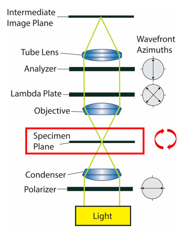
Polarized light microscopes are conventional standard microscopes equipped with two additional polarizing elements (the polarizer and the analyzer) that permit specimen examination under polarized light (see Fig. 1.).
The simplest polarized light microscope is a bright-field microscope (basic optical microscope which produces a dark image against a bright background) with a rotating stage and plane-polarizing filters placed below (the first polarizer, also called simply polarizer) and above (the second polarizer, also called analyzer) the specimen.
Light waves from the microscope lamp have randomly oriented planes of electric field oscillation (also called planes of polarization). The only light in which the electric field oscillates in parallel to the so-called privileged direction of the polarizer can pass through and are called plane-polarized light. When the privileged direction of the analyzer is oriented at right to that of the polarizer (i.e. they are at 90°C to each other), the field of view is in cross-polarized light, and the background will be dark. Image contrast arises from the interaction of plane-polarized light with a birefringent (or doubly-refracting) specimen to produce two individual wave components that are each polarized in mutually perpendicular planes. The velocities of these components, which are termed the ordinary and the extraordinary wavefronts, are different and vary with the propagation direction through the specimen. After transmitting the specimen, the light components become out of phase, but are recombined with constructive and destructive interference when they pass through the analyzer. [14]
The polarized light microscope allows observing samples that are visible primarily due to their optically anisotropic character. Anisotropic materials have optical properties that vary with the orientation of the incident light with respect to the crystallographic axis. [4] Such materials have more than one refractive index (for example, azurite, cinnabar, etc.) and with such microscopes will appear bright and distinctly coloured against the black background. However, these are polarization colours (not actual sample colours) and are related to the refractive indices and thickness of the sample. [15] In cross-polarized light, isotropic materials (e.g. gasses, liquids, resins, unstressed glasses, cubic crystals that have only one refractive index) will not be visible. [4,15]
The polarizing microscope is very useful for the pigments studies of paintings because most pigments have crystalline nature. Also, most pigments can be distinguished immediately from the binding media. [15] Furthermore, PLM can provide information about pigment’s refractive index, particle size and its distribution, colour, shape, pleochroism, optical properties, and morphology. [16] Pleochroism is a phenomenon when a crystal changes its colour or colour intensity when rotated in polarized light. This phenomenon happens as different crystallographic orientations absorb light differently. One good example is cobalt phosphate (cobalt violet), where experiments with PLM have shown colours from pink to lilac to orange-yellow. More about PLM analysis of pigments with multiple examples can be found in the book “Pigment Compendium, A Dictionary and Optical Microscopy of Historical Pigments” by Nicholas Eastaugh et al. [17].
PLM enables examining different fibres quite efficiently. PLM can reveal fibre’s surface characteristics, diameter, and optical properties like refractive index and birefringence (this means, can discriminate identical fibres chemically but have morphological differences).
The birefringence (Δn) can be calculated with the following formula: Δn = nII + n⊥ (nII – refractive index parallel to the axis along the fibre; n⊥ – refractive index perpendicular to the axis across the fibre). [4]
Polarized light microscopy is also widely used in archaeology for identifying, e.g. different plant remains (e.g. plant tissues, phytoliths, starch grains, etc.) absorbed into and attached on the surface of archaeological artefacts or found in sediments. In many cases, their shapes are diagnostic and helpful in identifying plants processed or consumed by ancient people. One of the most important characteristics of starch is that it glows under polarized microscopy and shows distinct x-shape images in the middle. This enables identifying starch-rich substances in ancient material. See Fig. 2.
 conventional optical and B) polarized light microscopies (photos by Dr Kristiina Johanson).")