Morphology of Nervous System
The spinal cord
The spinal cord is a cylindrical part of the CNS, approximately 40-45 cm long and 1.5 cm wide, situated in the vertebral canal. The spinal cord consists of the gray and white matter.
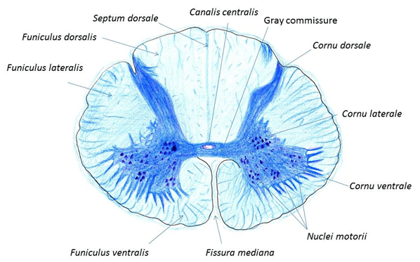
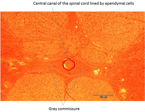
The gray matter is situated in the interior of the spinal cord and is divided into anterior, posterior and lateral columns (horns; cornu ventrale, cornu dorsale, cornu laterale), all three joined by the central gray commissure, where the central canal is located. The central canal is lined by a single layer of columnar ependymal cells – a sub-type of supportive neuroglial cells of the central nervous system. The gray matter contains neuron cell bodies and dendrites. Functionally related groups of nerve cell bodies are called nuclei.
The white matter consists of ascending and descending nerve fibers grouped into the anterior, lateral and posterior funiculi. The posterior funiculi are separated by posterior median septum and the anterior funiculi by anterior median fissure.

The spinal cord


